|
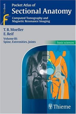
Pocket Atlas of Sectional Anatomy, Computed Tomography and Magnetic Resonance Imaging, Volume 3: Spine, Extremities, JointsTorsten Bert Moeller
יצא לאור ע"י הוצאת Thieme Medical Publishers,
שפת הספר: אנגלית |
|
תקציר הספר
Known to radiologists around the world for its superior illustrations and practical features, the "Pocket Atlas of Sectional Anatomy" now reflects the very latest in state-of-the-art imaging technology. In the classroom and the clinic, this compact book is a highly specialized navigational tool for radiologists on the road to diagnostic success.
Highlights of Volume III: Spine, Extremities, Joints
- All new superior CT and MRI images created using state-of-the-art equipment.
- More than 400 illustrations.
- Expanded and updated coverage of the structures of the arm, shoulder, elbow, hand, leg, hip, knee, foot, and spine.
- Each image of the spine, extremities, and joints is placed next to a clear, full-color diagram.
- Consistent color coding, making it easy to identify individual structures across several slices.
- Didactic approach and consistent format throughout -- an ideal study aid.
- Concise , easy-to-read labels and sophisticated diagrams.
- A handy size and format -- it fits in your pocket!
Vol 3: Spine, Extremities, Joints and its companion books -- Volume 1: Head and Neck and Volume 2: Chest and Abdomen -- comprise a must-have resource for radiologists of all levels.
פרסומת
לקט ספרים מאת Torsten Bert Moeller
לצפיה ברשימה המלאה, עבור לדף הסופר של Torsten Bert Moeller
©2006-2023 לה"ו בחזקת חברת סימניה - המלצות ספרים אישיות בע"מ
צור קשר |
חנויות ספרים |
ספרים משומשים |
מחפש בנרות |
ספרים שכתבתי |
תנאי שימוש |
פרסם בסימניה |
מפת האתר |
מדף גדול מדף קטן |
חיפוש ספרים

