|
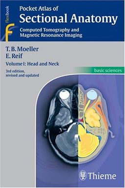
Pocket Atlas of Sectional Anatomy, Computed Tomography and Magnetic Resonance Imaing, Vol. 1: Head and NeckTorsten Bert Moeller
יצא לאור ע"י הוצאת Thieme,
שפת הספר: אנגלית |
|
תקציר הספר
Now with all new images!
Known to radiologists around the world for its superior illustrations and practical features, the "Pocket Atlas of Sectional Anatomy" now reflects the very latest in state-of-the-art imaging technology. In the classroom and the clinic, this compact book acts as a highly specialized navigational tool for radiologists on the road to diagnostic success.
Highlights of Volume 1: Head and Neck:
- All new CT and MRI images of the highest quality presented alongside brilliant full-color drawings.
- More than 413 illustrations.
- Even more slices per examination and more comprehensive coverage.
- Didactic approach and consistent format and throughout -- one slice per page.
- Concise, easy-to-read labeling on all figures -- a perfect balance of text and image
- Color-coded, schematic diagrams to indicate the level of the each section
-Sectional enlargements for a more detailed classification of the anatomic structure in question.
- The same handy size and format -- it fits in your pocket!
Vol. 1: Head and Neck and its companion books -- Volume 2: Chest and Abdomen and Volume 3: Spine, Extremities, Joints -- comprise a must-have resource for radiologists of all levels.
פרסומת
לקט ספרים מאת Torsten Bert Moeller
לצפיה ברשימה המלאה, עבור לדף הסופר של Torsten Bert Moeller
©2006-2023 לה"ו בחזקת חברת סימניה - המלצות ספרים אישיות בע"מ
צור קשר |
חנויות ספרים |
ספרים משומשים |
מחפש בנרות |
ספרים שכתבתי |
תנאי שימוש |
פרסם בסימניה |
מפת האתר |
מדף גדול מדף קטן |
חיפוש ספרים

